

Modern technology
Precise measurements are the basis for choosing the right treatment. At X-Vision we combine advanced diagnostics with modern laser and surgical technology for SMILE Pro, FS-LASIK, RLE lens exchange, ICL and cataract surgery. We have invested in modern equipment for both diagnostics and surgery, because the precision of the assessment determines how well the treatment can be planned for your eyes in particular. It is one of the reasons we call our practice a "refractive center of excellence".
We treat 13-14 eyes per day
Over 30 years of experience
Modern equipment
6-year Peace of Mind Guarantee
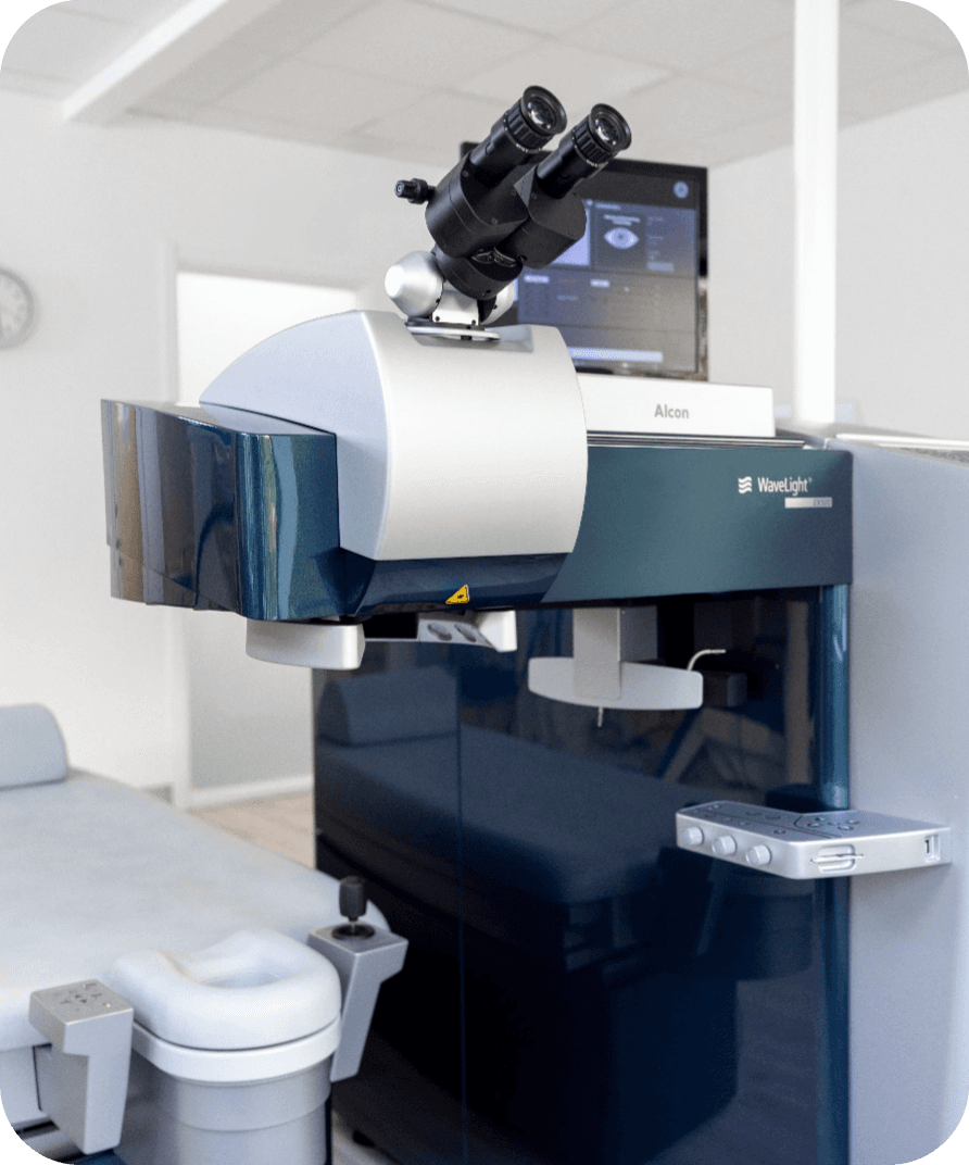
WaveLight EX500
The WaveLight EX500 excimer laser is FDA-approved in the USA and works at a repetition rate of 500 Hz, which keeps treatment times short. It has a high-precision eye tracking system, iris recognition, and can be paired with the Vario Topolyzer to perform Contoura LASIK. Together these features reduce fixation fatigue, limit corneal dehydration and support accurate centring during laser ablation. Combined with the VisuMax femtosecond laser, we use the WaveLight EX500 for bladeless LASIK.
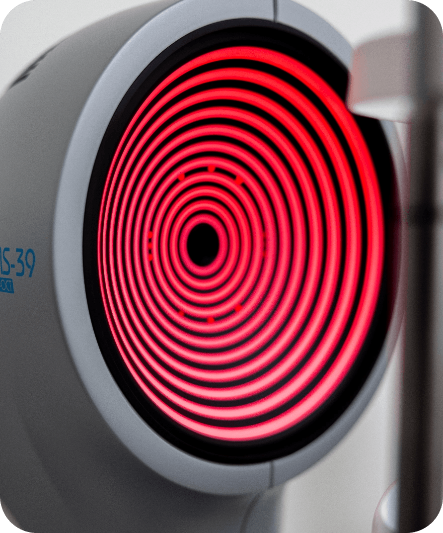
WaveLight Vario Topolyzer
The WaveLight Vario Topolyzer is an advanced device for corneal mapping, used to perform Topography-Guided Custom Ablation Treatment (T-CAT) or Contoura LASIK. By collecting data from 22,000 points on the cornea, the Vario Topolyzer generates a high-resolution image of the cornea and detects small irregularities known as higher-order aberrations. This information is passed to the WaveLight EX500 excimer laser to plan an individually tailored LASIK procedure.
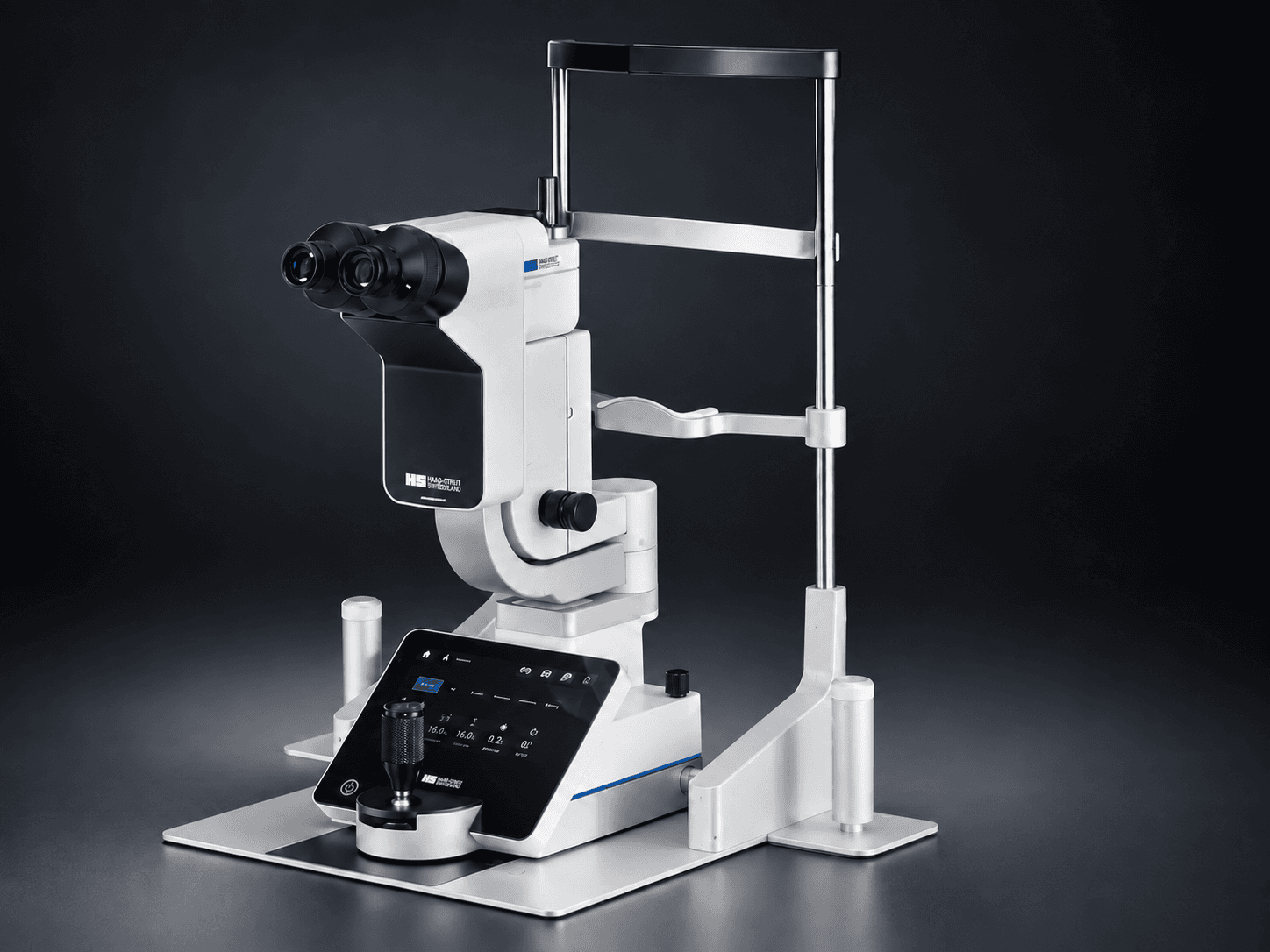
Elara 900 – slit lamp with image documentation
The slit lamp is where every examination begins. Under high magnification we assess the cornea, the anterior chamber, the iris, the lens and the tear film.
The Elara 900 has Haag-Streit optics and LED illumination with preset examination modes, so the same light and the same section can be reproduced at your next visit. That makes comparison over time reliable.
High-resolution images and video are captured in the lamp itself and stored in your record, so we can show you what we see – before and after a treatment, or while following a change in the cornea. An applanation tonometer for measuring eye pressure sits on the same instrument.
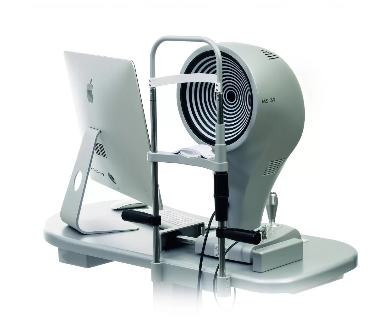
Epithelial mapping with the MS-39
MS-39 uses OCT (optical coherence tomography) to build a three-dimensional map of epithelial thickness, something not all corneal topographers can provide. Epithelial measurement is decisive for detecting early signs of keratoconus, individualising our laser treatments and understanding corneal remodelling after previous surgery.
MS-39 combines Scheimpflug + Placido + OCT, which gives a more complete picture of the whole cornea: the anterior and posterior surfaces, the stromal structure and pachymetry.
MS-39 maps both the anterior and posterior corneal surfaces and total thickness (pachymetry) more precisely, which matters for ectasia assessment, pre-operative screening (for every kind of vision correction) and follow-up after surgery.
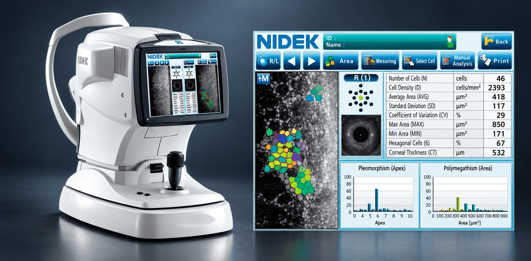
Nidek CEM-530 – the corneal endothelium
The CEM-530 photographs the endothelium, the innermost cell layer of the cornea. The cells are counted automatically, giving cell density, the variation in cell size and the proportion of hexagonal cells – centrally, paracentrally and peripherally, without the instrument touching the eye.
The endothelium pumps fluid out of the cornea and keeps it clear, and these cells are not replaced. That is why we measure it before ICL lens implants and lens exchange, in anyone who has worn contact lenses for many years, in Fuchs' endothelial dystrophy and after previous eye surgery.
Osiris – the eye's total optics
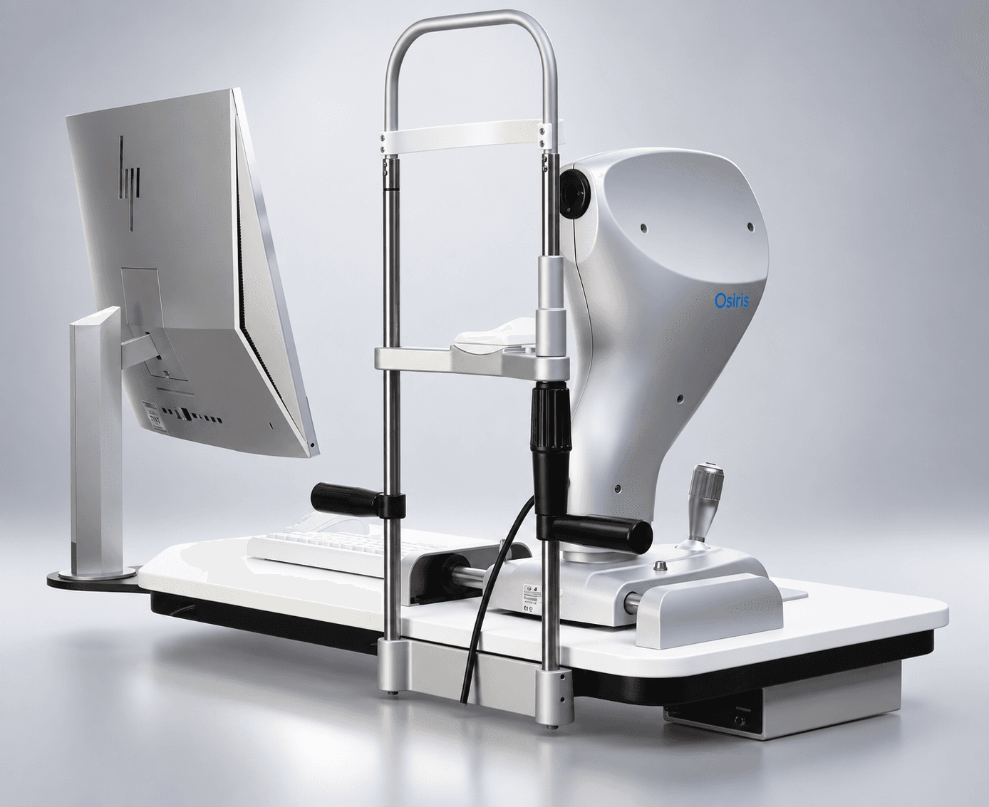
Osiris measures the total optics of the eye, not just the cornea. The aberrometer is built on pyramidal wavefront sensing, which samples more points than traditional Hartmann-Shack sensors and therefore also copes with irregular corneas.
Cornea or internal optics? We can separate how much of the refractive error sits in the cornea from how much comes from the internal optics of the eye. That bears on whether laser, a lens or a combination is the right route for you.
Higher-order aberrations and pupil size under different lighting are measured, which informs our assessment of the risk of glare and halos – and our choice of premium lens.
Together with the MS-39, Osiris gives a combined picture of the cornea and of the eye's total optics.
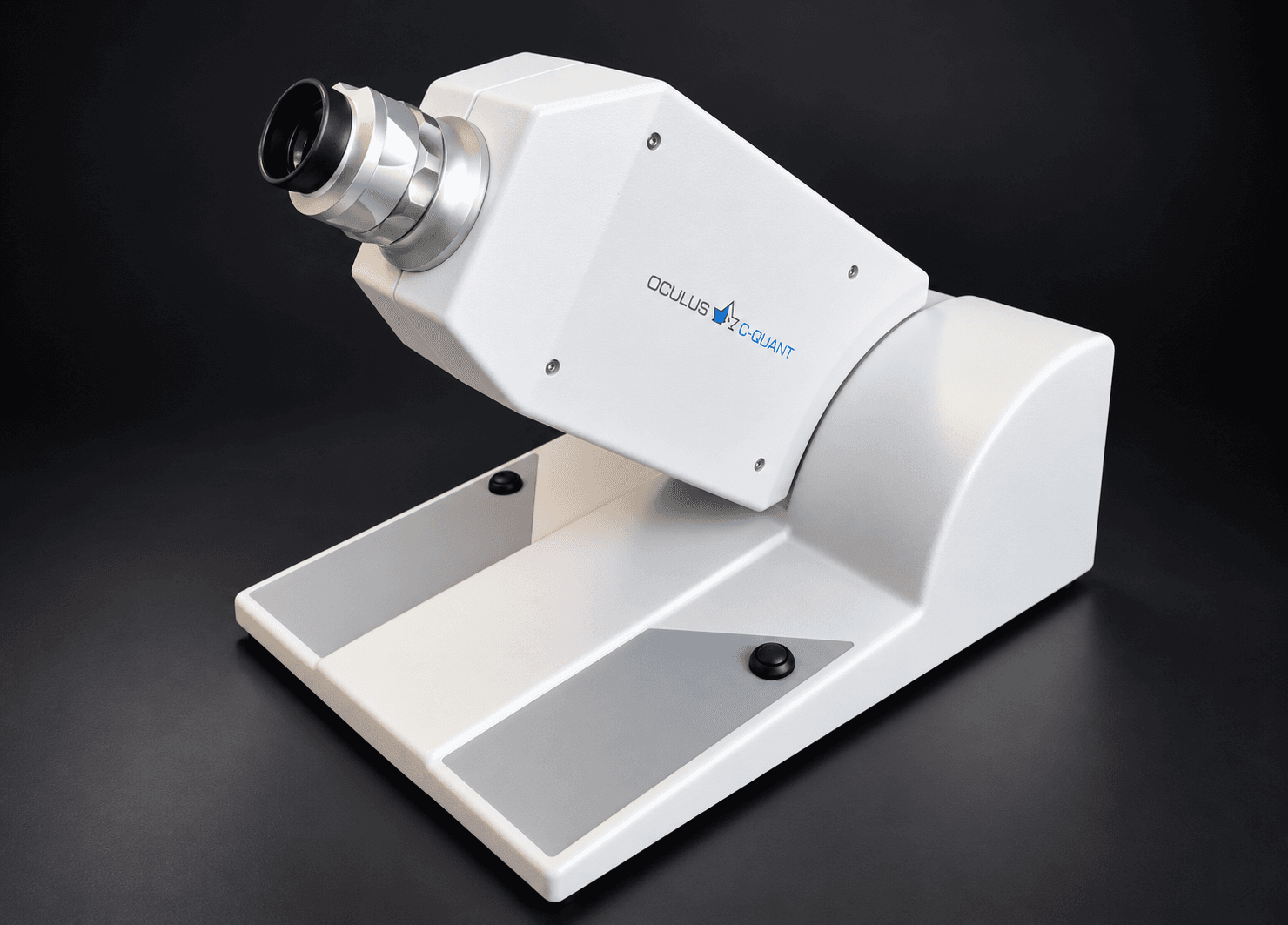
C-Quant – straylight and glare
Letters on a chart do not tell the whole story about how you actually see.
The C-Quant measures straylight: how much light is scattered inside the eye and settles as a veil over the image. This is the part of vision you notice with oncoming headlights at night, against the light and in fog – and it can be clearly reduced despite full visual acuity.
The measurement is psychophysical. You compare two halves of a flickering field, and the result is a value that can be followed over time. We use it in early cataract, with opacities in the lens or cornea, and when someone describes symptoms that acuity testing does not show.
HD Analyzer – the optical quality of the eye
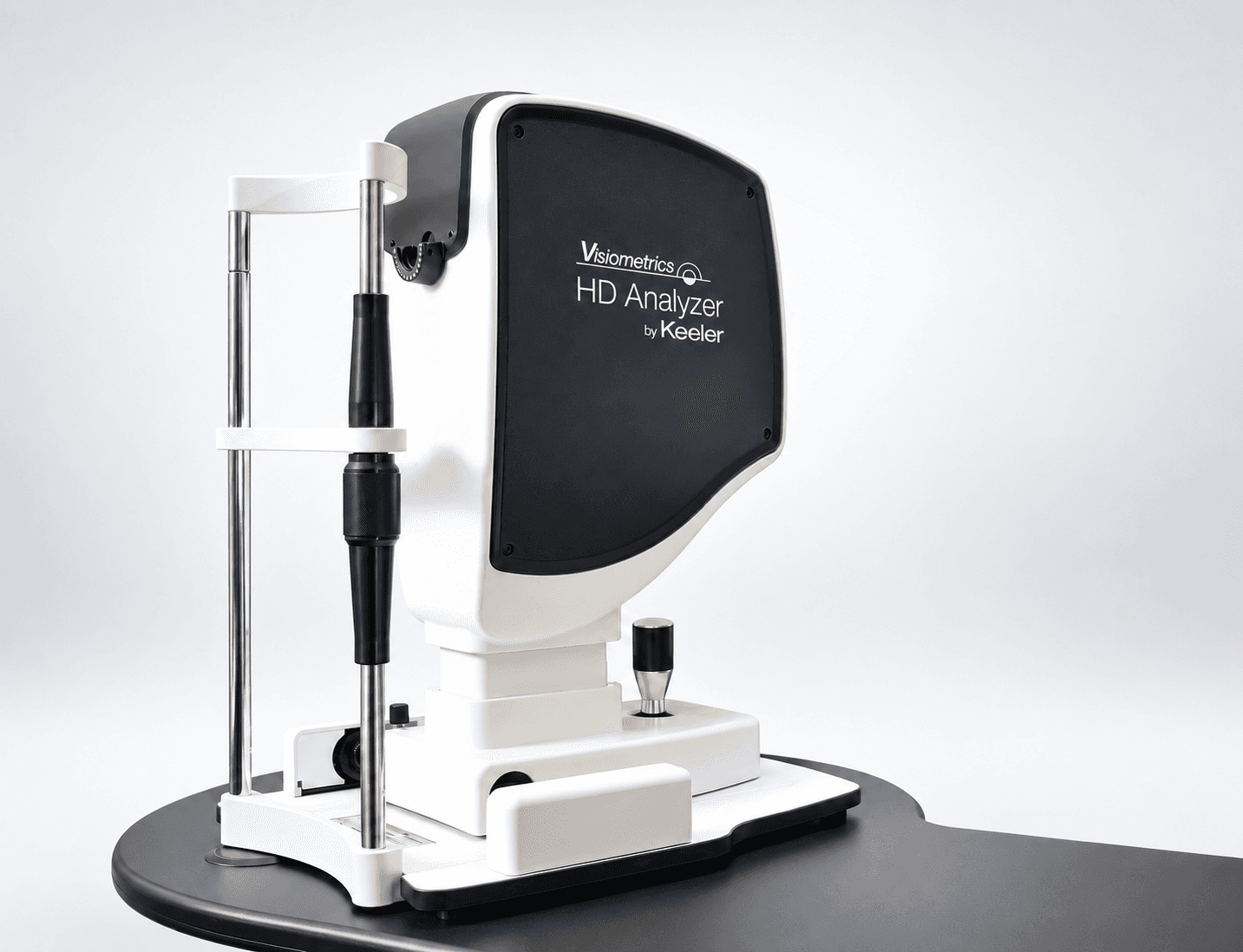
The HD Analyzer measures optical quality using the double-pass technique: a point of light is sent into the eye, and the image of that point on its way back carries everything the eye has done to the light going in and coming out.
From this we obtain the Objective Scatter Index (OSI), a measure of light scatter throughout the eye, and we can see how the tear film affects vision between blinks. The measurement requires no judgement from you.
It is useful in dry eye, in the work-up before vision correction, and when we want to establish how much of a visual complaint comes from the optics of the eye and how much has another cause. Together with the C-Quant and Osiris it gives a combined picture of visual quality.
Luxor Revalia
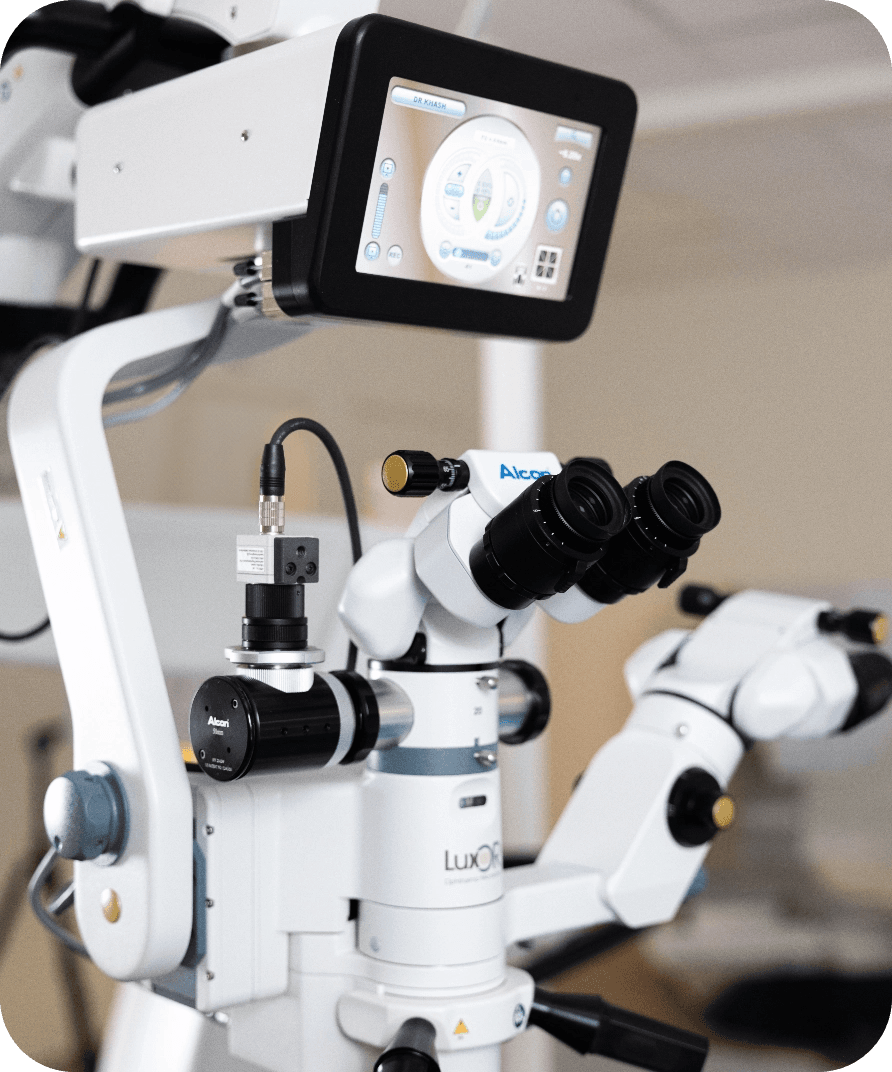
The surgical microscope we use for ICL lens implants, RLE lens exchange and cataract surgery. Combined with ORA, the Luxor Revalia provides evaluation, planning and real-time measurement during surgery, supporting precise placement of the lens.
IOL Master 700
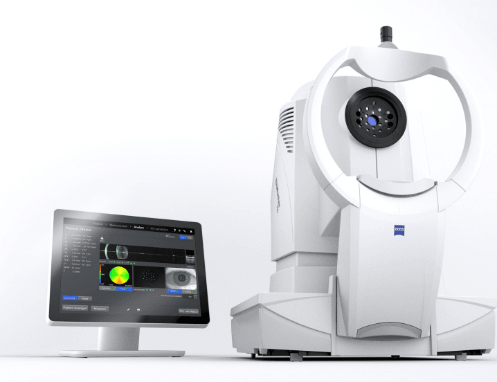
The ZEISS IOL Master 700 is an advanced optical biometer used before lens exchange surgery to measure the anatomy of the eye with high precision. Swept-source OCT visualises the eye's entire optical system, which ensures correct measurements and reveals irregularities. The examination is fast, entirely contact-free and possible in 99% of cases, even with a dense cataract, which reduces the need for ultrasound. Total Keratometry measures both the anterior and the posterior corneal surface, while the Barrett Suite gives more accurate lens calculations. Integration with the ZEISS Cataract Suite enables markerless surgery for precise placement of toric lenses. The result is high accuracy in the lens calculation, a shorter examination and greater patient safety.
Eyestar 900 – biometry from cornea to retina
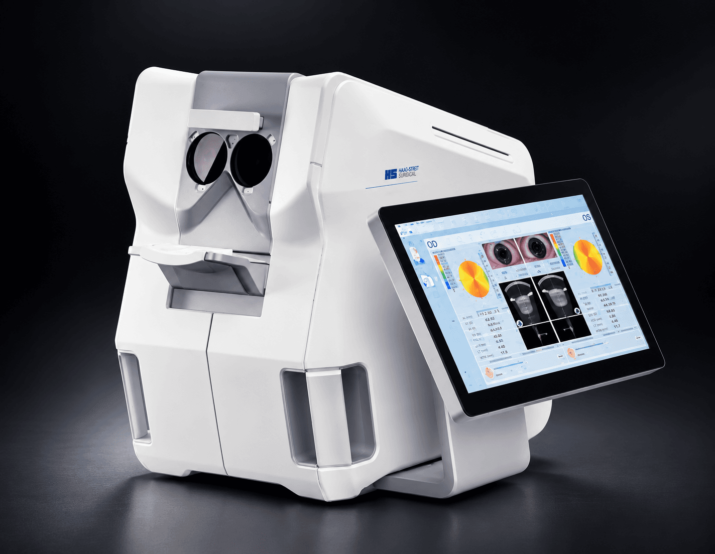
The Eyestar 900 is a biometer built on swept-source OCT. It measures the whole eye in a single examination – axial length, corneal curvature in two zones, the anterior and posterior corneal surfaces, thickness (pachymetry), anterior chamber depth and the position of the lens – and shows cross-sectional images of what was measured.
Before lens exchange and cataract surgery it gives us a second, independent set of biometry alongside the IOL Master 700, so the calculations can be compared before we settle on a lens and its power. Topography out to 12 mm and lens tilt analysis inform our planning for toric and multifocal lenses.
Topcon DRI OCT Triton – retina and choroid
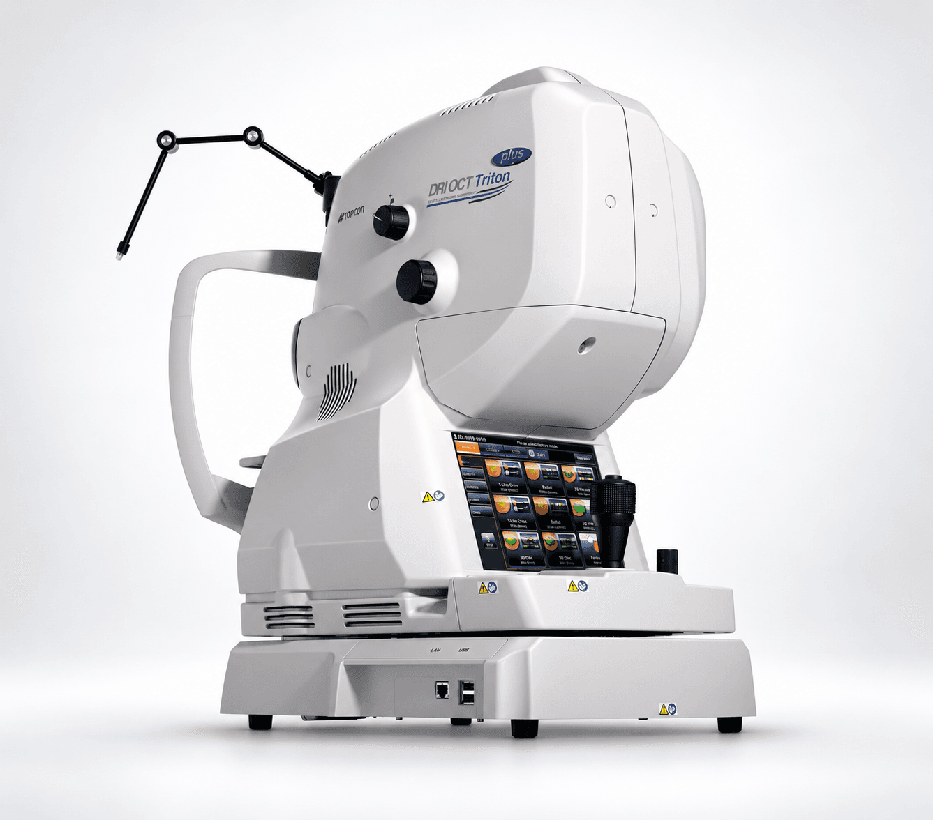
The DRI OCT Triton is a swept-source OCT for the retina. It works at 1050 nm at 100,000 A-scans per second, which gives deeper penetration – down into the choroid – and images that hold up even once the lens has started to cloud with cataract.
The instrument also performs OCT angiography, mapping the blood vessels of the retina and choroid without an injection of contrast, and captures colour images of the retina in the same session.
We use it for the macula, in diabetes, before lens exchange and cataract surgery, and whenever a loss of vision needs to be explained behind the lens. It also lets us separate an optical problem from a retinal one before planning vision correction.
VisuMax 800
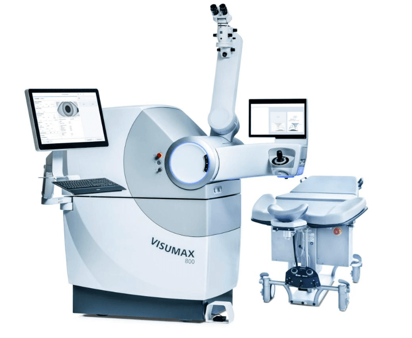
VisuMax is the femtosecond laser we use to create a LASIK flap, and the VisuMax 800 is the only laser that can perform SMILE Pro laser eye surgery. It is made by Zeiss, known for its precision optics. The ZEISS VisuMax 800 works at a laser frequency of up to 2 MHz (2000 kHz), and with SMILE Pro the laser step itself can take under 10 seconds per eye, depending on the treatment. The low pressure on the eye and the short laser time make the procedure quick and comfortable.
C-Eye – corneal cross-linking
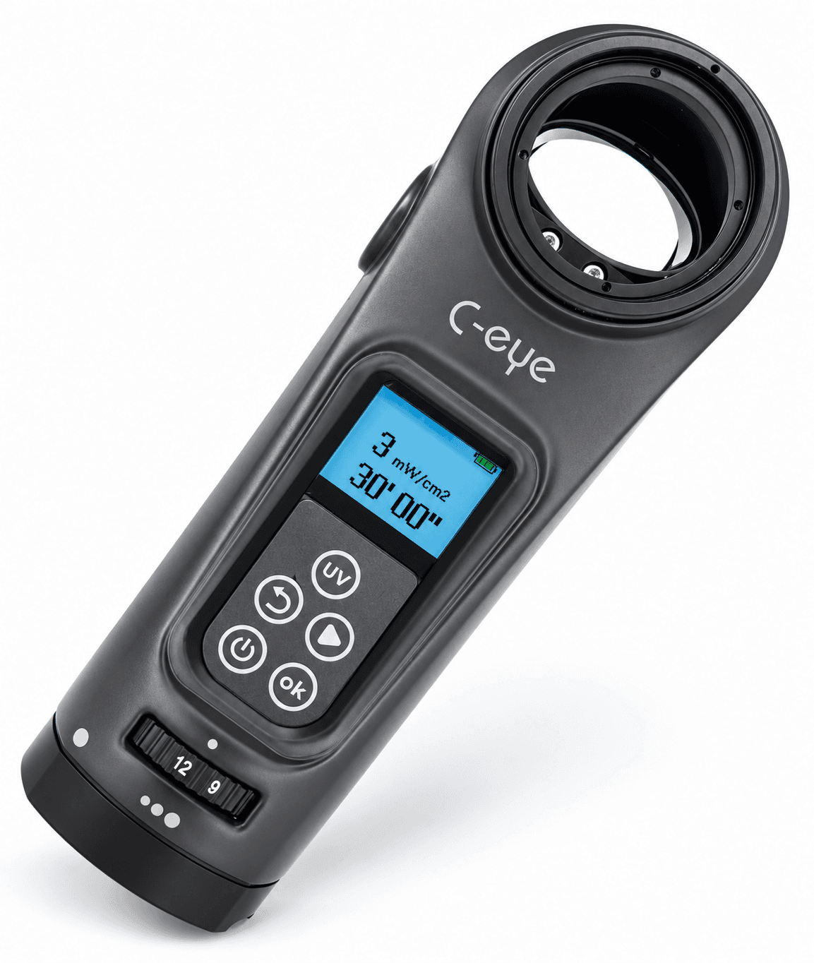
C-Eye is our system for corneal cross-linking (CXL) in keratoconus. Established protocols are preset – from the classic Dresden protocol to accelerated, high-fluence and sub-400 protocols for thinner corneas.
Irradiance, time and total dose are set and documented for every treatment, and it can be given with or without removing the epithelium. The same system is used for PACK-CXL, cross-linking in corneal infections. Read more about cross-linking
Nidek YC-200 S plus – YAG and SLT
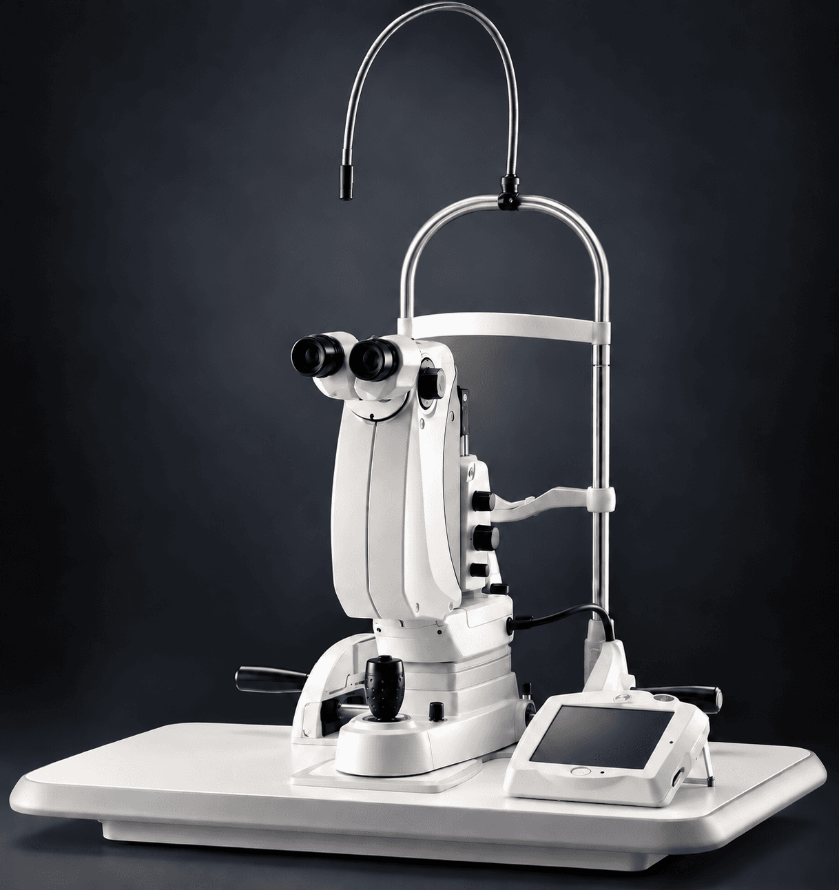
The YC-200 S plus is a combined laser. In YAG mode it is used for posterior capsule opacification (capsulotomy) – when the capsule behind an implanted lens has clouded – and for iridotomy. In SLT mode we treat raised eye pressure in open-angle glaucoma.
The optics have extended depth of focus and LED illumination, and the cone angle of the beam is designed so that energy density at the cornea is lower than in older designs. The plasma threshold is low, so treatment can be carried out with less energy. In SLT mode the treated areas are displayed as we go, so the dose is spread evenly around the angle.
Both treatments are done under anaesthetic drops and take a few minutes. Together with the Ellex Ultra Q Reflex, which we use for vitreolysis, this covers the laser treatments that may be needed after lens surgery or in glaucoma.
Ellex Ultra Q Reflex
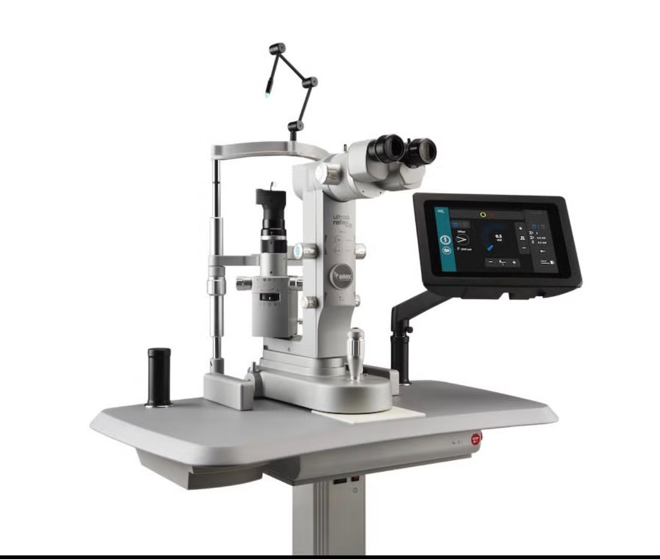
The Ellex Ultra Q Reflex is the YAG laser we use for vitreolysis – laser treatment of vitreous floaters – and for YAG capsulotomy after cataract surgery.
Its optics are designed for work in the vitreous: coaxial illumination and a converging beam make it easier to see floaters in the mid-vitreous and to place the focal point with clearance from both the lens and the retina.
Whether vitreolysis is suitable is decided at the examination. The position, size and distance of the floater from the retina govern what can be treated – and not every floater is suitable for laser.
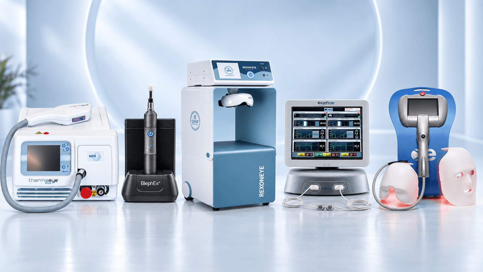
Equipment for treating dry eyes
Dry eye has several different causes, so treatment has to be aimed at whatever is actually producing the symptoms. We keep a range of techniques at the clinic so that the method can follow what the examination shows.
ThermaEye Plus (IPL) – intense pulsed light for inflammation of the lid margin and meibomian gland dysfunction.
Eye-Light (IPL and LLLT) – pulsed light and low-level light therapy. These can be given in the same session or separately.
REXON-Eye (QMR) – quantum molecular resonance, acting on the whole ocular surface.
LipiFlow – warms the meibomian glands from the inner surface of the lid and empties them with pulsating pressure.
MGX – manual expression of the meibomian glands.
BlephEx – cleaning of the lid margin in blepharitis and Demodex.
Which treatment suits you is decided at the examination, and the treatments can be given individually. Read more about dry eyes
Contact us – we'll help you find the right treatment
Which treatment suits your eyes? Tell us briefly what you would like help with and we will get in touch. Our experienced specialists answer your questions and guide you to the treatment or examination that fits your needs.
X-Vision Ögonklinik
Hyllie Stationstorg 2
215 32 Malmö